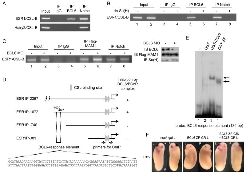
Figure 6.
The mechanisms by which BCL6 shuts down the expression of selected Notch target genes.
A: ChIP assays were performed with nuclear extracts from stage 25 embryos using α-BCL6 antibody, α-Notch1 antibody or mouse IgG. Mouse IgG was used for a mock ChIP assay. B: 2 ng dn-Su(H) was injected into two-cell stage embryos and nuclear extracts were isolated at stage 10 for ChIP assays. C: BCL6 MO or/and 1 ng Flag-MAM1 was injected into two-cell stage embryos and nuclear extracts were isolated at stage 10 for ChIP assays. The levels of input proteins were confirmed by immunoblotting. D: Deleted fragments of X. tropicalis ESR1 gene were linked to the luciferase reporter. Numbers indicate the position of nucleotides from the initiation site. E: Incubation of GST-BCL6 or GST-ZF with a probe corresponding to a 134-bp (-1030/-897) element yielded one distinct retarded band indicated by an arrow. F: 2 ng BCL6 ZF-GR or/and mBCL6-GR was injected for each experiment. The injected side is indicated by L (left) beside the names of injected samples. L: left, R: right, a: anterior, p: posterior.

