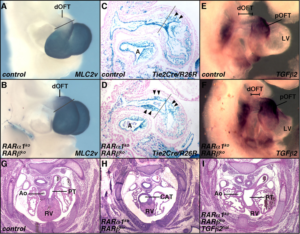
Fig. 3.
OFT axial misspecification and elevated TGFβ cause CAT. A–B. Ectopic expression of the proximal marker MLC2v in the distal OFT of RAR mutant embryos at E10.5. C–D. Endocardial mesenchyme (arrowheads) in the distal OFT of mutant embryos at E10.5, visualized by Tie2Cre/R26R. Gray lines in A–D are positioned at the 90° bend between the proximal and distal segments of the OFT. E–F. Elevated Tgfb2 expression in the distal OFT in mutants at E9.75. G–I. Rescue of septation defects in E14.5 RAR mutants by reduced Tgfb2 gene dosage: a normal control embryo (G), a RARα1/RARβ mutant with CAT (H), and a RARα1/RARβ mutant also heterozygous for Tgfb2 (I). In rescued embryos, OFT septation occurs but both outflow vessels originate from the right ventricle (the RV source of the ascending aorta is not seen in this panel). Ao, ascending aorta; PT, pulmonary trunk. See also Fig. S3.

