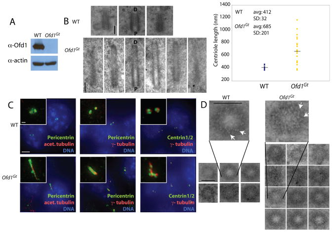
Figure 1. Ofd1 is essential for centriole length control.
(A) Immunoblot of cell lysate supernatants from wild type (WT) and Ofd1Gt cells. 15 μg protein loaded per lane. (B) Longitudinal TEM sections of WT and Ofd1Gt cell centrioles. Long centrioles (defined as > 600 nm) are seen in 35% of Ofd1Gt cells. P, proximal end and D, distal end of centriole. Graph shows centriole length data, collected from 9 WT and 23 Ofd1Gt centrioles. Each measured centriole was from a distinct cell. (C) Representative fluorescence micrographs of WT and Ofd1Gt cells showing centrosomes (Pericentrin and γ-tubulin), centrioles (Centrin and acetylated tubulin), and DNA (DAPI). (D) Transverse TEM sections of WT and Ofd1Gt cell centrioles. White arrows indicate triplet microtubules. Normal length centrioles are contained within a maximum of 8–10 transverse sections, whereas long centrioles span more than 10 sections. Scale bars indicate 200 nm (TEM), 5 μm, and 1 μm (inset). See also Figure S1.

