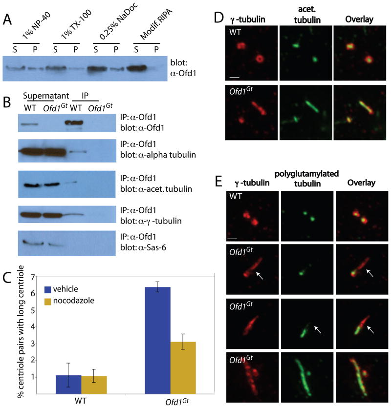
Figure 3. Ofd1 complexes contain centriolar microtubule components and control centriole microtubule stability.
(A) Immunoblot showing Ofd1 (detected with an Ofd1 antibody) in the supernatant (S) and pellet (P) of WT cells lysed with various detergents. (B) Immunoblots of Ofd1 complexes immunoprecipitated from WT or Ofd1Gt cell supernatant with an Ofd1 antibody. (C) Graph indicating percent of long centrioles in WT and Ofd1Gt cells. Cells were treated with nocodazole, fixed and stained for α- and γ-tubulin. γ-tubulin foci more than twice as long as they were wide were counted as long centrioles. Because immunofluorescent (IF) microscopy has lower resolution than TEM, a smaller percent of Ofd1Gt centrioles appeared long when assessed by IF (6–10% by IF versus 35% by TEM). (D) WT and Ofd1Gt cells stained for centrosomes (γ-tubulin) and acetylated tubulin. (E) WT and Ofd1Gt cells stained for centrosomes (γ-tubulin) and polyglutamylated tubulin (GT335). Arrows indicate areas of reduced or absent polyglutamylation. Scale bars indicate 1μm.

