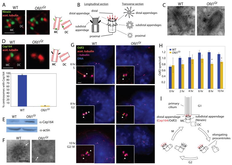
Figure 5. Ofd1 is essential for distal appendage formation.
(A) Centrioles (acetylated tubulin) and mother centriole subdistal appendages (Ninein) of WT and Ofd1Gt cells. (B) Diagram depicting the TEM appearance of a mother centriole in transverse and longitudinal views. (C) TEM longitudinal views of WT and Ofd1Gt centrioles. Arrows indicate subdistal appendages. (D) Centrioles (acetylated tubulin) and mother centriole distal appendages (Cep164) of WT and Ofd1Gt cells. Graph shows percent of centrosome pairs showing Cep164 localization in WT and Ofd1Gt cells. (E) Immunblot showing Cep164 in the supernatants of WT and Ofd1Gt cell lysates. 20 μg protein loaded per lane. (F) TEM transverse views of WT and Ofd1Gt centrioles. Arrows indicate distal appendages. (Full serial reconstructions are included in Figure S4). (G) Centrioles and cilia (acetylated tubulin), centriole appendages (Odf2), centrosomes (γ-tubulin), and DNA (DAPI) of WT and Ofd1Gt cells at the indicated time after release from cell synchronization block. Arrowheads indicate centrioles positive for Odf2. (H) Quantification of Odf2 foci per cell in WT and Ofd1Gt cells at the indicated time after release from cell synchronization block. Asterisks indicate p< 0.05. (I) Diagram showing centriole duplication and maturation in coordination with the cell cycle. MC, mother centriole. DC, daughter centriole. Scale bar indicates 200 nm (TEM) and 1 μm. See also Figure S4.

