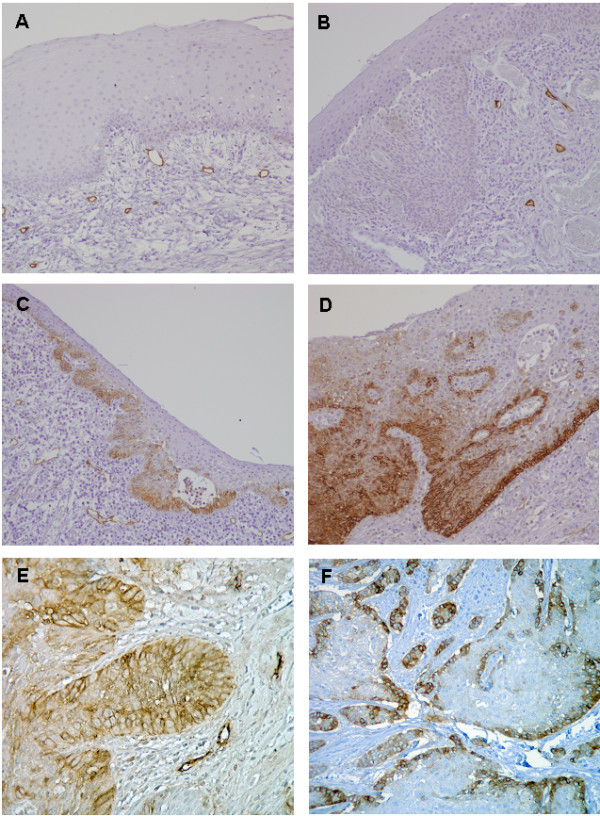
Figure 1.

Immunohistochemical analysis of podoplanin expression. Representative examples of podoplanin expression in normal laryngeal epithelium (A), dysplastic epithelium with negative podoplanin staining (B), dysplastic epithelium with positive podoplanin staining scored as 2 (C) and 3 (D), laryngeal carcinomas showing diffuse podoplanin expression (E) and focal expression (F).
