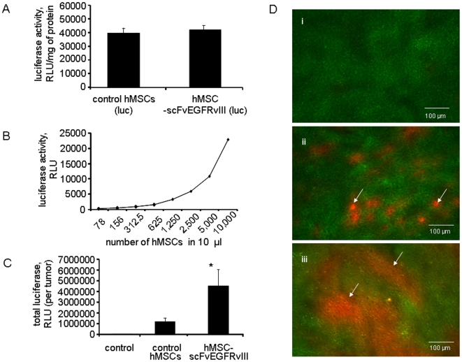
Figure 2. hMSC-scFvEGFRvIII presence in U87-EGFRvIII flank xenografts in athymic mice.
A. hMSCs-scFv and control hMSCs were nucleofected with pGL4.14 plasmid encoding luciferase gene and stable population was selected with hygromycin. Control and scFvEGFRvIII expressing hMSC cells show equal expression of luciferase. Data presented as mean ± SD, n = 3. B. Titration of hMSC expressing luciferase. Sensitivity of assay. C. Presence of hMSC-vector and hMSCscFvEGFRvIII in the s.c. flank U87-EGFRvIII xenograft at day 25 either alone or after co-injection of control hMSC- vector or hMSC-scFvEGFRvIII cells (n = 8 in each group). Luciferase assay. Data presented as mean ± SD, * p<0.05. D. Microphotograph of 10 µm tissue sections from s.c. xenograft of (i) GFP expressing U87-EGFRvIII cells alone and either (ii) with control hMSC-vector or (iii) hMSC-scFvEGFRvIII cells at day 21. Arrows indicate the presence of hMSC expressing RFP.

