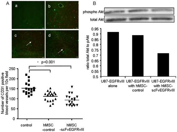
Figure 5. Brain tumor vascularization in the presence of hMSC-scFvEGFRvIII and pAkt expression.
A. Tissue section (10 µm) from flash frozen brains carrying tumors were stained for mouse CD31, (a) negative control, U87-EGFRvIII GFP expressing tumor; (b) CD31 positive blood vessels in U87-EGFRvIII GFP expressing tumor; (c) CD31 positive blood vessels in U87-EGFRvIII GFP expressing tumor co-injected with control hMSCs; (d) CD31 positive blood vessels in U87-EGFRvIII GFP expressing tumor co-injected with hMSC-scFvEGFRvIII. Arrows show CD31 positive blood vessels. The number of CD31 positive blood vessels was calculated in each field (n = 18) (magnification 10×) and data summarized in the graph. * p<0.001. B. Expression of pAkt in glioma cells sorted from 4 week old tumors established with U87-EGFRvIII alone or with control or scFvEGFRvIII expressing hMSCs. Representative Western Blot from two independent experiments is shown.

