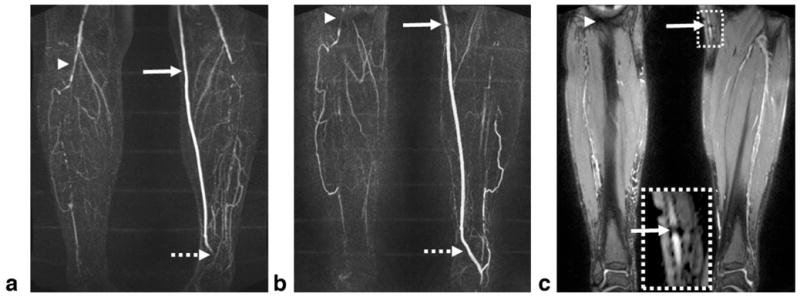
FIG. 10.

Subtraction MIP images for CE-MRA (a) and NC-MRA (b) in a 65-year-old male patient with known PAD. Both techniques demonstrate significant bilateral calf artery occlusion. The large collateral vessels are not well delineated on the CE-MRA image due to incomplete arterial enhancement (right leg) or venous overlay (left leg). The patent bypass graft (CE-MRA image) appears to have a stenosis (solid arrow) on the NC-MRA image, which is most likely due to the adjacent metallic clip–induced signal loss in the bright-artery scan (c). The distal part of the bypass graft (dashed arrow) is not shown on the CE-MRA image since it is not included in the imaging volume. At the edge of the FOV (arrowhead), artery delineation is compromised by banding artifacts when using the bSSFP-based NC-MRA technique. The CE-MRA and NC-MRA scans have slightly different FOV as the imaging session was interrupted at request of the patient.
