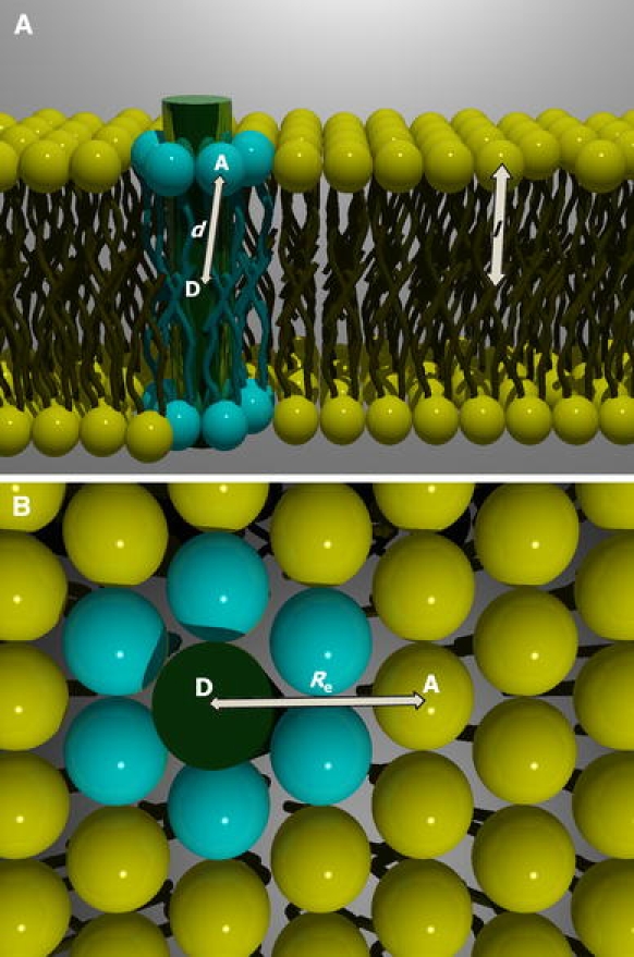
Fig. 3.

Molecular model for the FRET analysis according to the model of Fernandes et al. (2004): (a) side view and (b) top view. Protein–lipid organization presents a hexagonal geometry. Donor fluorophore from the mutant protein is located in the center of the bilayer, whereas the acceptors are distributed in the bilayer surface. Two different environments are available for the labeled lipids (acceptors): the annular shell surrounding the protein and the bulk lipid. Energy transfer to acceptors in direct contact with the protein has a rate coefficient dependent on the distance between donor and annular acceptor (Eq. 5). Energy transfer toward acceptors in the bulk lipid is given by Eq. 6
