
Fig. 1.
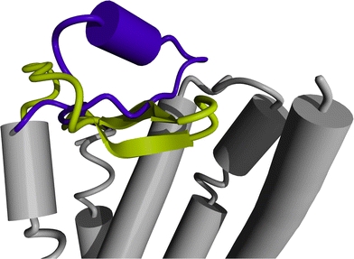
The most striking difference between the crystal structures of rhodopsin (PDBid 1f88, Palczewski et al. 2006) and the beta-2 adrenergic receptor (PDBid 2rh1, Cherezov et al. 2007) concerns the structure and location of the extracellular loop between helix IV and V. The loop IV–V in rhodopsin forms a β-sheet that folds into the binding pocket (yellow), whereas loop IV–V in the beta-2 adrenergic receptor forms an α-helix and extends towards the extracellular environment (purple)