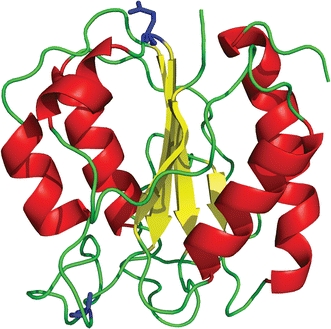
Fig. 1.

Schematic diagram of native C69A flavodoxin from A. vinelandii (pdb ID 1YOB (Alagaratnam et al. 2005)). The protein contains a parallel β-sheet surrounded by α-helices at either side of the sheet. Residues Ala-69 and Gln-48 are shown in ball-and-stick representation (coloured blue). The FMN cofactor is not shown
