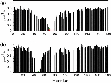
Fig. 6.

PRE of amide protons of WTMTSL apoflavodoxin and Q48CMTSL apoflavodoxin, which are both unfolded in 6.0 M GuHCl. 1H–15N HSQC spectra of both protein variants with the spin-label either in the paramagnetic or diamagnetic state were recorded at 25°C. Subsequently, the ratio of the intensities of cross peak maxima (I para/I dia) of backbone amides was determined. Shown are I para/I dia of a WTMTSL apoflavodoxin and b Q48CMTSL apoflavodoxin. Green dots highlight the positions of the spin-label. Red bars in (a) highlight residues for which no cross peaks are visible in the HSQC spectrum of the protein with paramagnetic spin-label, but for which cross peaks are observed when the spin-label is diamagnetic
