Fig. 6.
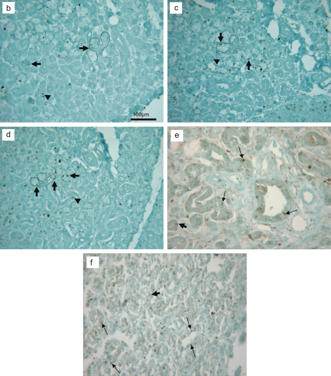
Cell proliferation in the deligated glands. a, number of ki-67 positive nuclei per field of view (×200 magnification) in the 2 weeks ligated plus 3 or 5 or 7 days deligated (3 days reg.; 5 days reg.; 7 days reg.) submandibular gland. 5 days deligated gland showed an increase in the number of proliferating cells (p<0.05, 3 observation in 5 rats, n=15) compared to the 3 days. b, c, d ki-67 immunostaining (counterstained with Light Green dye) in the 2 weeks ligated plus 3 or 5 or 7 days deligated respectively, showing mostly proliferating acinar cells (arrows), also present at the end of the branched structures (dashed silhouettes), and occasionally ductal cells (arrowheads). e, f ki-67 immunostaining of normal adult and ligated rat submandibular gland respectively. Normal gland (e) showed proliferation of ductal cells (arrow) and occasionally acinar cells (thick arrow). The ligated gland (f) revealed proliferation of ductal cells (arrows) and some non-parenchymal cells (thick arrow). All tissue sections were counterstained with Light Green.


