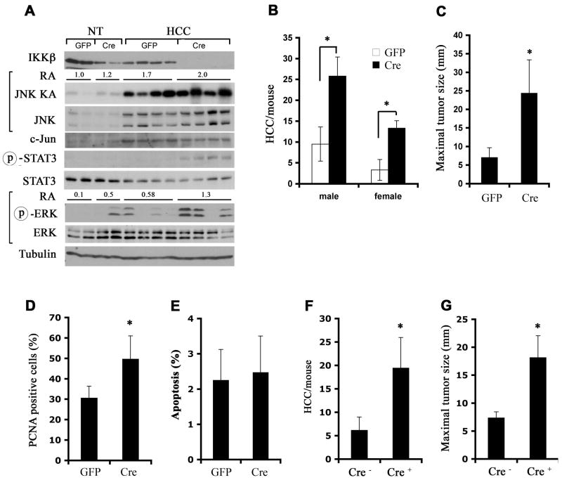
Figure 2. IKKβ deletion after initiation enhances HCC formation and growth.
(A–C) Ikkβf/f male pups were DEN initiated and used as hepatocyte donors into MUP-uPA mice. IKKβ in transplanted hepatocytes was deleted 1 month later by injection of Adv-Cre. Adv-GFP was used as a control. Mice were sacrificed 4 months later and whole cell lysates were prepared from dissected HCCs and surrounding non-tumor livers (NT).
(A) Lysates were gel separated and immunoblotted with the indicated antibodies or used for measurement of JNK kinase activity by immunecomplex kinase assay. Relative JNK kinase activity (KA) and ERK phosphorylation in the different samples were determined by densitometry and the average relative activities (RA) for each group of samples are indicated.
(B) HCCs per liver were counted and (C) maximal tumor size was measured. n = 7~10 for each group; *: p < 0.05.
(D, E) Tumor cell proliferation and apoptosis were determined by PCNA (D) and TUNEL (E) staining, respectively, of paraffin embedded liver sections (n = 10 each group; *: p < 0.05).
(F, G) Ikkβf/f (Cre−) and Ikkβf/f/Mx1-Cre (Cre+) DEN-initiated males were used as hepatocyte donors to male MUP-uPA recipients. IKKβ deletion was accomplished by poly(IC) injection 1 month after transplantation. HCC multiplicity (F) and maximal sizes (G) were determined 4 months later (n = 10 each group; *: p < 0.01).
Please also see Figure S2.

