Figure 5.
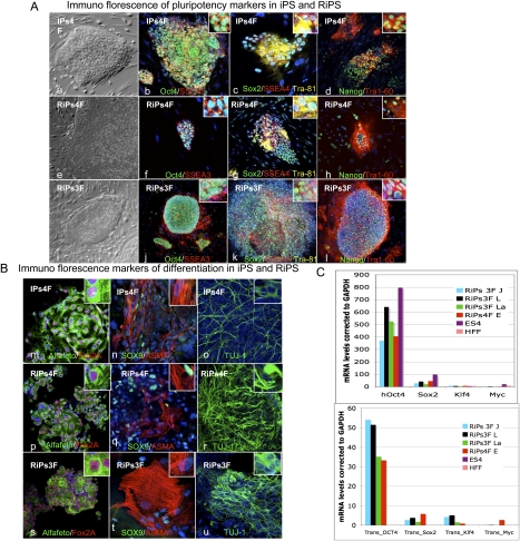
Characterization of RiPs. (A, panels a–l) Phase contrast and immunostaining of iPSCs (one clone) and RiPs (two clones) for pluripotency markers Oct4, SSEA3, Sox2, SSEA4, Tra-81, Nanog, and Tra-60. (B, panels m–u) Immunostaining of differentiation markers following 3 wk of general differentiation of iPSC and RiP clones (gelatin with 20% FCS medium) for Alfafeto protein, Fox2A, Sox9, Alfa smooth muscle (ASMA), and TUJ1. (C) Real-time PCR of transgenes and pluripotency marker expression in RiPs compared with hESCs and somatic cells.

