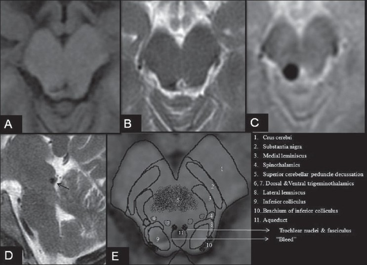
Figure 1.

MRI brain in a patient with isolated acute left trochlear nerve palsy. Axial T1 SE (A), axial T2 FSE (B), Axial T2*GRE (C) and Sagittal T2WI(D) showing a hypointense lesion in the right tectal region at the level of the inferior colliculus with intense blooming on gradient imaging without any flow voids. E, schematic representation of the anatomical structures of the midbrain at the level of the inferior colliculus overlapped on Figure A. Note: Increased hypointensity of a paramagnetic substance such as blood on T2*GRE (gradient) imaging is termed as blooming
