Figure 1.

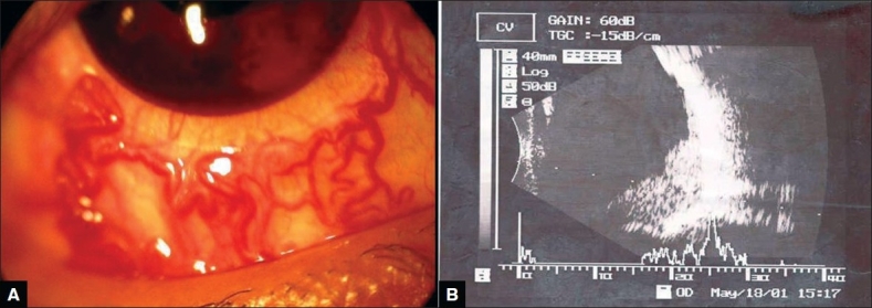
(A) Racemose dilatation of conjunctival blood vessels and note cosmetic contact lens in position. (B) USG picture showing mass lesion in inferior and infero-temporal quadrant

(A) Racemose dilatation of conjunctival blood vessels and note cosmetic contact lens in position. (B) USG picture showing mass lesion in inferior and infero-temporal quadrant