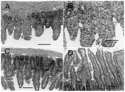
Figure 4.
Photomicrographs of mouse distal ileum 7 days p.i. with T. gondii. (A and B) Parental C57BL/6 mouse showing acute necrosis of small bowel villi with acute inflammatory infiltration and ulceration. (A) Bar equals 100 μM. (B) Bar equals 200 μM. (C) Parental mouse treated with aminoguanidine showing preservation of small bowel epithelium. Bar equals 100 μM. (D) iNOS−/− mouse with normal small bowel epithelium. Bar equals 75 μM.

