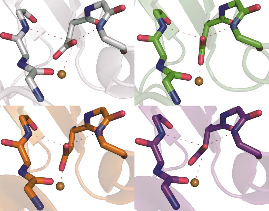
Figure 2. The position of D112 shifts among the proteins, leading to variations in hydrogen bonding to the carboxylate.
Secondary coordination spheres of Cu(II) in C112D (a, 1.9 Å, PDBID: 3FQY), C112D/M121L (b, 2.1 Å, PDBID: 3FPY), C112D/M121F (c, 1.9 Å, PDBID: 3FQ2), and C112D/M121I (d, 1.9 Å, PDBID: 3FQ1) azurins are highlighted with bond distances shown in Å for heteroatoms involved in the hydrogen bonding “rack” network of the wild-type protein. Oxygen atoms are red; nitrogen atoms are blue.

