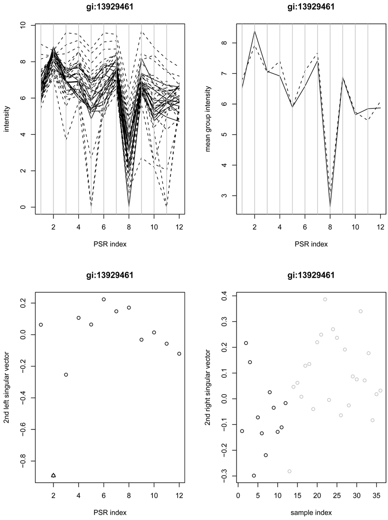
Figure 3.
Example gene gi:13929461 is detected by the SVD method as the “stage 2”, but missed by ANOVA. The left upper panel contains the plot of intensities of individual samples versus PSRs (solid-normal; dashed-cancer); The right upper panel contains the average intensities of normal samples (solid) and cancer samples (dashed) versus PSRs; The left lower panel contains the plot of the second left singular vector along with the PSRs (an outlying PSR indicated by triangle); The right lower panel contains the second right singular vector versus samples (black-normal; grey-cancer).

