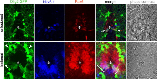
Figure 2.
Evidence for a pMN-equivalent zone in the ependymal layer of the adult spinal cord. Spinal cross-sections at the level of the central canal (asterisks) are shown. In the unlesioned (arrows) and lesioned spinal cord (brackets) Pax6/Tg(olig2:egfp)/Nkx6.1 coexpressing ependymoradial glial cells are present. Expression of all markers is increased close to a spinal lesion site at 2 weeks postlesion. Arrowheads, Oligodendrocytes. Scale bar, 20 μm.

