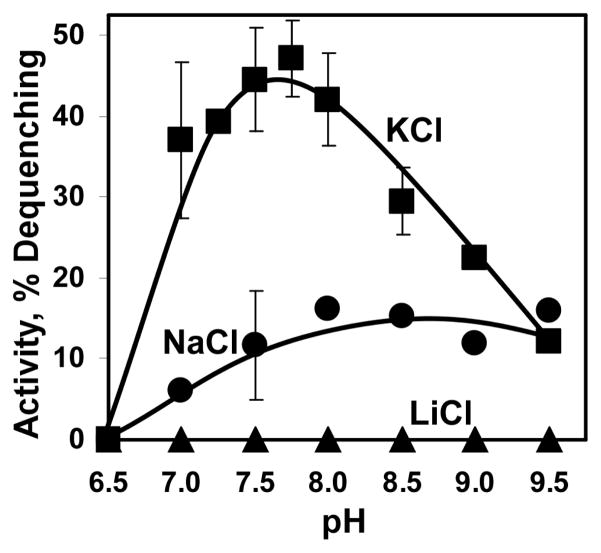
FIGURE 3.
pH profile of Vc-NhaP2 activity. Inside-out membrane vesicles were isolated from TO114 cells transformed with pVc-NhaP2 or “empty” pBAD24 and assayed with the specified salt in standard choline chloride buffer adjusted to the indicated pH with 50 mM BTP-HCl. In each case, residual non-specific activity measured in “empty” vesicles was substracted from that registered in Vc-NhaP2-containing vesicles and the resulting Vc-NhaP2-dependent activity was plotted as a function of pH. All other conditions as in Fig. 2. Plotted are the averages of six measurements (carried out in duplicate with three separate isolations of vesicles).

