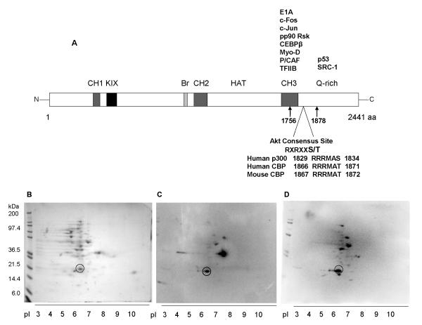
Fig. 1.
Experimental pI of T7-tagged fragment of CBP spanning the CH3 domain and partial Q-domain, CBP CH3. (A) Diagram of different domains of CBP, including partial list of known interacting proteins at the C-terminus. Location of the Akt consensus site within the cloned fragment of mouse CBP is indicated. The analogous Akt consensus sites at human CBP and p300 are also shown. (B) Two-dimensional gel electrophoresis of CBP CH3 lysates and subsequent detection of resolved proteins via Coomassie blue staining and (C) parallel Western blotting against T7 tag and (D) Akt substrate motif.

