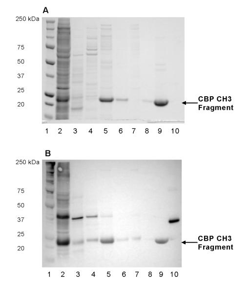
Fig. 4.
Progress of purification of CBP CH3 proteins as monitored by (A) Coomassie blue staining and (B) Western blotting against T7 tag. Arrows indicate the position of observed MW of T7-tagged CBP CH3 proteins. Lane 1: MW markers; lane 2: cell lysate; lane 3: Post HA A11-A13 fractions; lane 4: Post HA G7-G14 fractions; lane 5: Post HA H-I fractions; lane 6: FT of Post HA H-I fractions; lane 7: Post SEC A5-A10 fractions; lane 8: FT of Post SEC A13-B12 fractions; lane 9: Post SEC A13-B12 concentrate; lane 10: T7-tagged positive control (31.1 kDa). HA, hydroxyapatite; SEC, Superdex 200; FT, flow-through.

