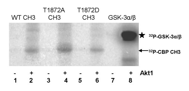
Fig. 8.
In vitro phosphorylation assays of CBP CH3 fragments and GSK-3α/β fusion protein in the absence and presence of Akt1. 32P-labeled proteins were identified in PVDF membrane by autoradiography. Arrow indicates the location of 32P-CBP CH3 fragments, while star indicates the location of 32P-GSK-3α/β.

