
Fig. 2.
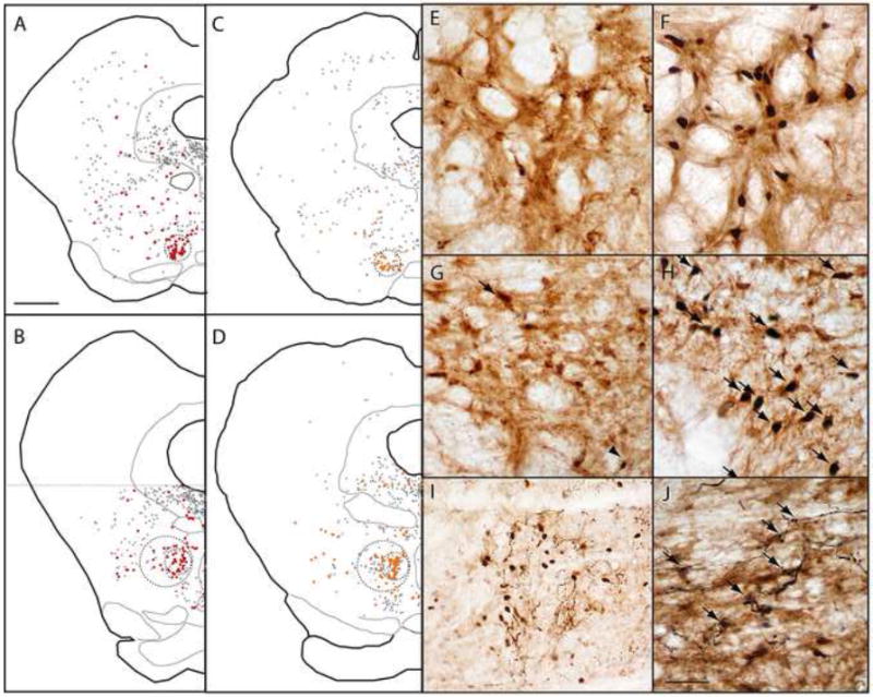
(A-D) After retrograde tracer injections into the VTA, retrogradely labeled neurons are clustered in the RMTg, but also found throughout the reticular formation (black and red filled symbols). Retrogradely labeled neurons co-expressing GAD67 (red symbols in A-B) are dense within the RMTg, as are retrogradely labeled neurons expressing shock-induced Fos (orange symbols in C-D). Colabeling in rostral (A, C) and caudal (B, D) levels, is particularly high within the central “core” of the RMTg (smaller dashed circles), is less dense in the RMTg “periphery” (larger dashed circles in B and D), and is uncommon outside the RMTg. Photomicrographs of the RMTg from unshocked (E) and shocked (F) rats show marked shock-induced Fos (black nuclei) in retrogradely labeled neurons (brown cytoplasmic label). Similarly, GAD67-immunoreactive neurons in the RMTg contain very little Fos in unshocked rats (G), but colocalize heavily with Fos after footshock presentation (H, arrows). After anterograde tracer injections into the medial portion of the lateral habenula (LHb), labeled fibers in the RMTg intermingle with shock-activated Fos nuclei (I), with less labeling where Fos nuclei are absent. In a separate case, labeled fibers arising from the LHb appose RMTg soma that are retrogradely labeled by a CTB injection into the VTA (J, arrows). Scalebar in A is 1mm (applies to A-D). Scalebar in J is 250μm (I) or 100μm (E-H, J).