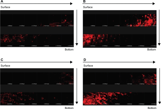
Figure 6.
Confocal laser scanning microscopic (CLSM) micrographs of nude mouse skin after the in vivo topical administration of sulforhodamine B (SRB) via the skin for 2 hours from 30% ethanol (control) without fluorescein isothiocyanate (FITC) loading (A) NLC-5P without FITC loading (B) 30% ethanol (control) with FITC loading (C) NLC-5P with FITC loading (D).
Notes: The skin specimen was viewed by CLSM at ∼10-μm increments through the Z-axis from skin surface. The arrows indicate the skin fragments from surface to bottom.

