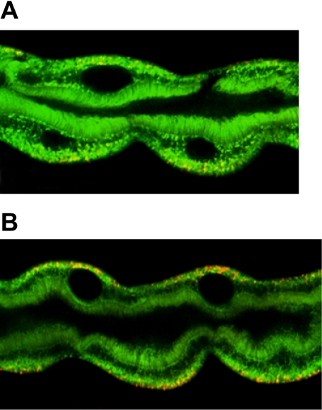
Fig. 5.
Mitochondria are overactive in sesB mutants. A, B: imaging of live tubules using mitochondrial potential-sensitive dyes. Activated mitochondria, in which the inner membrane potential is hyperpolarized, accumulate red fluorescence with JC-1 (48). A: mitochondrial activity in tubules from hs-GAL4 flies. B: note the increase in the red fluorescence compared with A in basal surface of tubule principal cells from hs-GAL4 > UAS-sesB RNAi flies.

