Fig. 5.
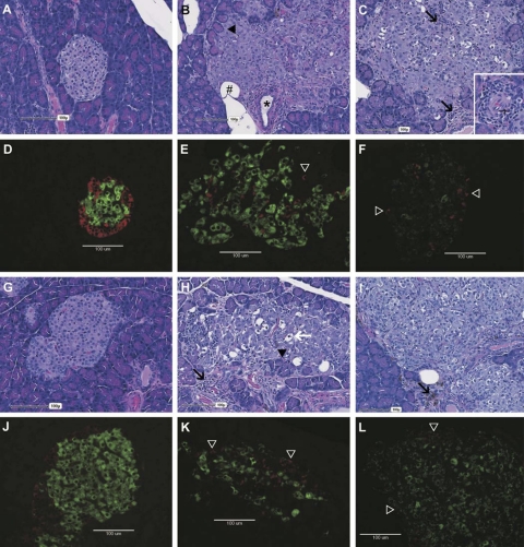
Representative hematoxylin and eosin (H & E)-stained histological and insulin and glucagon immunofluorescent sections from female (A–F) and male (G–L) rats. Wild-type DR.+/+ (left); DR.lepr−/lepr− (center), and DR.lepr−/lepr−,Gimap5−/Gimap5− (right) rats are shown. Age and no. of days with diabetes: DR.+/+ control rat at 180 days of age (A, D, G, and J); nondiabetic female DR.lepr−/lepr− rat at 180 days of age (B and E); 113-day diabetic DR.lepr−/lepr− male rat at 180 days of age (H and K); 27-day diabetic DR.lepr−/lepr−,Gimap5−/Gimap5− rat at 150 days of age (C and F), and 36-day diabetic DR.lepr−/lepr−,Gimap5−/Gimap5− rats at 81 days of age (I and L). Wild-type islets are clearly demarcated from exocrine acinar cells with central insulin-producing β-cells (green) surrounded by a ring of glucagon-producing α-cells (red) in females (A and D) and males (G and J). In contrast, DR.lepr−/lepr− islets in females (B and E) and males (H and K) are enlarged with an irregular shape, encroachment, and occasional entrapment of acinar exocrine cells (black arrowheads). Immunofluorescent sections stained for insulin and glucagon confirm that the enlarged islets noted on H & E-stained sections are composed of abundant β-cells and disrupted α-cells (E and K, open arrowheads) with multifocal black regions within the islets likely representing entrapped acinar cells (B and H, black arrowheads), lipocytes (B, #), ducts (B, *) or regions of inflammatory (H, black arrows) or degenerate islet cells characterized by enlarged cells with condensed to pyknotic nuclei, occasional karyorrhexis, and vacuolated cytoplasm (H, white arrow). Note the relative increase in degenerate cells within the male DR.lepr−/lepr− islet compared with the female DR.lepr−/lepr− islet. Enlarged islets were also noted in DR.lepr−/lepr−,Gimap5−/Gimap5− double congenic rats (C and F, female; I and L, male), immunostaining again confirming that enlarged islets contained abundant β-cells and disrupted α-cells (F and L, open arrowheads). Note the perivascular, periductular, and intraislet mixed inflammation (C, inset and I, black arrow) composed of lymphocytes and macrophages, occasionally hemosiderin laden (I, arrow), eosinophils (C, inset and black arrows), and neutrophils.

