
Figure 2.
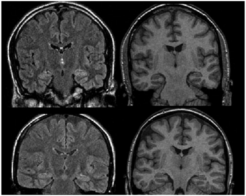
Examples of subtle abnormalities appreciated on MRI re-review. Upper panel shows FLAIR (left) and T1-SPGR (right) images of subtle left hippocampal signal abnormality without atrophy. Lower panel shows FLAIR (left) and T1-SPGR (right) images of subtle left hippocampal atrophy without signal abnormality.
Epilepsia © ILAE