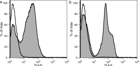
Fig. 5.
Flow cytometry detection. (a) Yeasts displaying HA–Fn3–c-myc (irrelevant Fn3 clone) were labeled with PBS (empty) or anti-HA goat IgG (shaded) followed by DyLight633-conjugated gI2.5.3T88I and analyzed by flow cytometry. (b) As in (a) except anti-c-myc rabbit IgG and rI4.5.5K27S/K56S were used. Note that the two peaks in the IgG-labeled samples correspond to cells with and without plasmid, which serves as an effective internal control.

