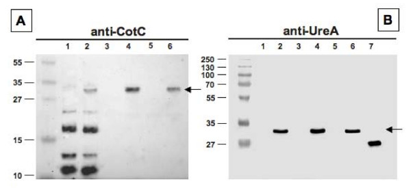
Figure 3.
Western blot analysis of Fusion 2 (CotC-UreA1) performed with anti-CotC (A) or anti-UreA (B) of spore coat proteins extracted from strains PY79 (lanes 1), KH10 (PY79 carrying cotC::ureA1) (lanes 2), RH101 (PY79 cotC::spc) (lanes 3), KH11 (PY79 cotC::spc cotC::ureA1) (lanes 4), RH209 (PY79 cotC::spc cotU::erm) (lanes 5) and KH12 (PY79 cotC::spc cotU::erm cotC::ureA1) (lanes 6). In both panels arrows point to fusion proteins. Purified UreA was run in lane 7 of panels B. Twenty five micrograms of total proteins were separated on either 15% (A) or 12% (B) polyacrylamide gels, electrotransferred to nitrocellulose membranes and reacted with primary antibodies and then with peroxidase-conjugated secondary antibodies and visualized by the enhanced chemiluminescence method.

