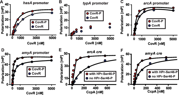
Figure 5. Recombinant CovR and CcpA bind to DNA from promoter regions of the same genes.
(A-D) Representative fluorescence polarization-based isotherms of unphosphorylated (blue circles) and phosphorylated CovR (red circles, CovR-P) binding to 1 nM of fluorescein-labeled DNA. Millipolarization units (mP) are plotted against the CovR concentration. (A) Recombinant CovR interaction with DNA from the hasA promoter (positive control). (B) CovR interaction with DNA from promoter of the non-CovR regulated gene typA (i.e. negative control). For (B) note linear increase in MP values with increasing CovR concentration indicating low affinity protein-DNA interaction. (C) Recombinant CovR interacting with DNA from the amino acid utilization gene arcA. (D) CovR interaction with DNA from the carbohydrate utilization gene amyA. CcpA interaction with arcA cre (E) and amyA cre (F) is shown with (red circles) and without (blue circles) the presence of 10 µm HPr-Ser46-P. For all panels experiments were done on at least three occasions, and lines indicate fit of binding data as described in Materials and Methods. hasA, hyaluronan synthase; typA, GTP-binding protein; arcA, arginine deminase; amyA, cyclomaltodextrin glucanotransferase.

