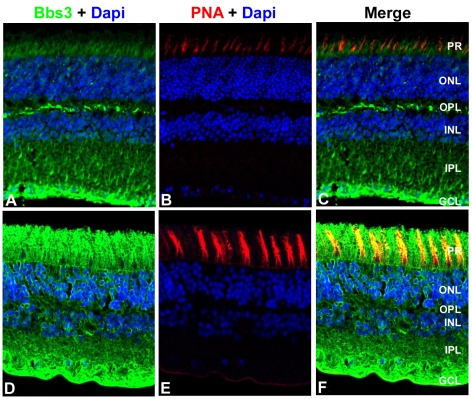
Figure 5. Localization of BBS3 in human and wild-type mouse retinas.
Immunohistochemistry triple labeling of cryosections taken from 8-month old wild-type mouse retinas (A–C) and human donor eyes (D–F). Localization of BBS3 (green) using an antibody generated against a central region of the mouse Bbs3 peptide, which recognizes both Bbs3 and Bbs3L (A,D). Peanut agglutinin (PNA, red) was used as a marker for cone outer segments (B,E). Nuclei were counterstained with DAPI (blue). Bbs3 was found robustly in the ganglion cell layer as well as the photoreceptor cell layer of human and mouse retinas. PR, photoreceptor; ONL, outer nuclear layer; OPL, outer plexiform layer; INL, inner nuclear layer; IPL, inner plexiform layer; GCL, ganglion cell layer.

