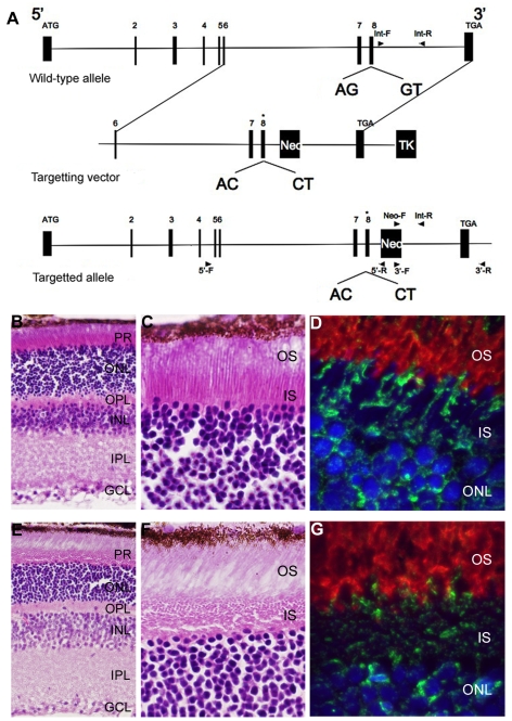
Figure 6. Generation and initial characterization of a Bbs3L mutant mice.
(A) Schematic for the targeted alteration of the splice donor and acceptor sites of exon 8 (asterisk) found in the Bbs3L transcript. Homologous recombination leads to the inclusion of these altered sites and the loss of Bbs3L. Hematoxylin/eosin staining of cryosections from 8-month old Bbs3L+/+ (B,C) and Bbs3L−/− (E,F) mouse retinas. Disruption of the normal photoreceptor architecture was observed in Bbs3L−/− mice. Immunohistochemistry analysis of cryosections from (D) wild-type and (G) targeted mutants retinas using the Bbs3 antibody (green) and rhodopsin (red), a marker for rod photoreceptor outer segments. To-Pro3 was used as a counterstain for nuclei. PR, photoreceptor; ONL, outer nuclear layer; OPL, outer plexiform layer; INL, inner nuclear layer; IPL, inner plexiform layer; GCL, ganglion cell layer.

