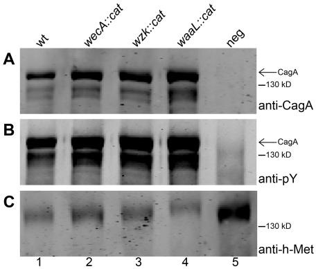
Figure 7. CagA translocation is functional in O antigen negative H. pylori strains.
Human gastric epithelial AGS cells were infected with H. pylori G27 (1) wild type, (2) wecA mutant, (3) wzk mutant, or (4) waaL mutant strains. (5) Non-infected AGS cells served as negative control. AGS membranes were examined for translocation and tyrosine phosphorylation of CagA by Western blotting using (A) anti-CagA antibody and (B) anti-phosphotyrosine (pY) antibody. (C) The host membrane marker h-Met was detected with an anti-h-Met antibody for a loading control. Arrows indicate full length phosphorylated CagA. A protein marker standard was included for reference.

