
Fig. 2.
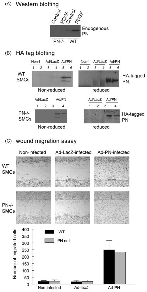
Effect of adenovirus-mediated overexpression of PN on SMC migration. (A) Western blotting demonstrated the expression of endogenous PN detected by using an anti-PN antibody in the conditioned medium obtained from quiescent cultures of WT and PN−/− SMCs, in the absence or presence of PDGF-BB (10 ng/ml) as indicated. (B) Western blotting demonstrated the expression of adenovirus-produced HA-tagged PN detected by using an anti-HA antibody either in the conditioned medium (lanes 1, 3 and 5) or the cellular lysates (lanes 2, 4 and 6) obtained from cultures of WT (top) and PN−/− (bottom) SMCs, non-infected (Non-I), infected with Ad-LacZ or Ad-PN as indicated. Under reducing conditions (with β-mercaptoethanol: β-ME), the PN migrates as a monomer (∼90 kDa). Under non-reducing conditions (without β-ME), the PN migrates as both a monomer and a dimer. (C) Migration by a scratch-wound assay showed that both WT (top) and PN−/− (bottom) SMCs infected by Ad-PN migrated into the wound area at 20 h after wounding, almost completely filling the wound space, whereas few non-infected or Ad-LacZ-infected cells migrated into the wound area. The lines delimit the initially wounded regions. The number of cells migrated into the wound space was measured from digital images. Results are representative of three separate experiments.