
Figure 6.
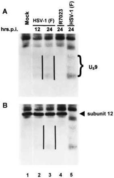
Immunoblot of infected cell proteins immunoprecipitated with a monoclonal antibody to proteasome, electrophoretically separated on a denaturing gel and allowed to react with a rabbit polyclonal antibody to US9 protein (A), photographed, and then allowed to react with a monoclonal antibody to subunit 12 of proteasome (B). The parallel lines are to the right of the US9 protein bands.