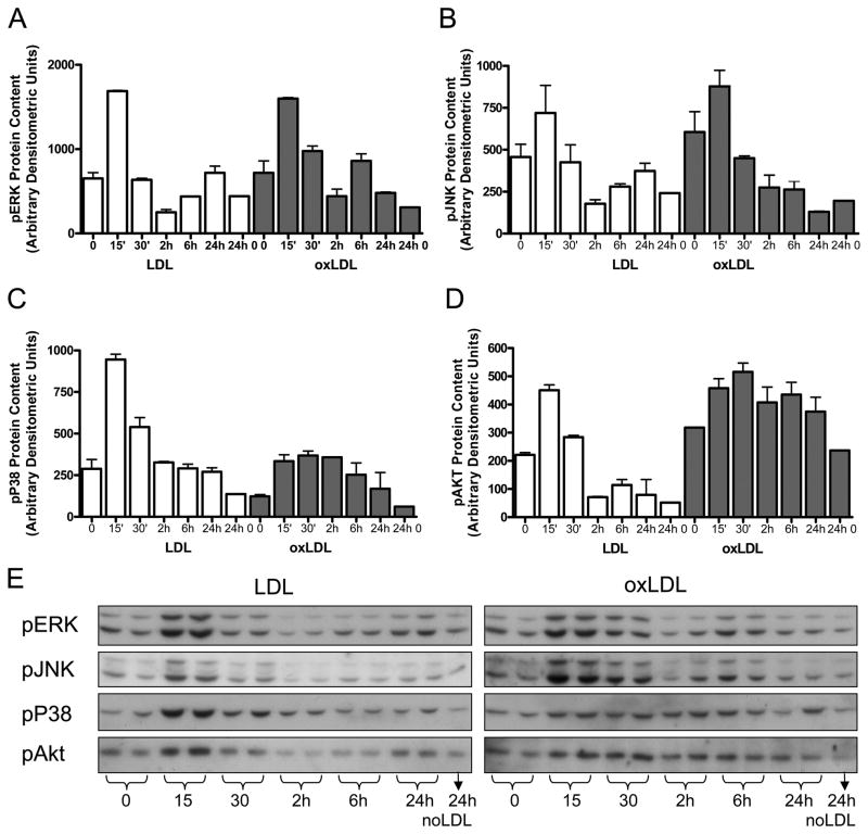
Figure 4.
Activation of MAPkinase and Akt pathways in primary culture rat VSMC after 15 minutes exposure to LDL or oxLDL (50 ug/ml). Phosphoprotein concentrations were quantified by densitometric analysis of Western blots with antibodies to (A) pERK, B) pJNK, (C) pP38, and D) pAkt. (E) Representative western blots with duplicate loading are shown. Values are mean of independent duplicate experiments and error bars represent the maximum value. Similar results were obtained in 2–4 separate experiments, each in duplicate or triplicate, for individual pathways.

