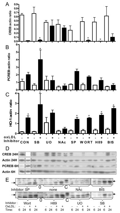
Figure 5.
Involvement of signaling pathways in the effects of oxLDL on primary culture rat VSMC. Cells were pretreated for 30 minutes with or without (CON) inhibitors of signaling pathways: SB-203580 (SB, 20 μM, p38 MAPK), SP-600125 (SP, 20μM, JNK MAPK), U-0126 (U0, 10 μM, ERK MAPK), Wortmannin (WORT, 100nM, PI3K), H89 (10 μM, PKA), Bisindolyl maleimide (BIS, 4μM, PKC), N-acetyl cysteine (NAC, 30mM, ROS), and then incubated with (+) or without (−) oxLDL (40 ug/ml) as indicated. CREB at 24 hours (A), PCREB at 6 hours (B) and HO-1 at 6 hours (C) were quantified and normalized to β-actin by densitometric analysis of Western blots. (D) Representative western blots are shown for CREB, PCREB, and β-actin at 6 and 24 hours. Individual portions of western blots were from two gels from a single experiment. A control extract was loaded on all gels to allow gel to gel comparison, and blots of comparable exposure are shown. (E) Representative western blots are shown for HO-1. Blot portions shown were from the same experiment, but multiple gels as in D. Note that inhibitor order is different from that shown in the graphs. Graphs represent data combined from three experiments, each with duplicate or triplicate independent samples. Because some experiments did not include all inhibitors, 3–7 independent samples are represented in each data point. Values are mean ± SD; p values for comparison to corresponding control are by ANOVA with Bonferroni correction for multiple comparisons. * p<0.05 vs. corresponding no oxLDL control.

