
Fig. 4.
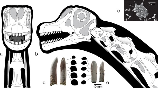
Reconstruction of the skull of A. mcintoshi based on holotypic and referred specimens (DINO 16488, 17848, 17849, 39727) in anterior (a) and left lateral (b) views. Computed tomography cross-section through the third cervical vertebra just posterior to the diapophysis (c) reveals camellate pneumaticity. Photographs of left premaxillary tooth 1 (d) and right dentary tooth 5 (e) in lingual, mesial, and cross-sectional views show differences in tooth shape. Note twisting of carina in the premaxillary tooth, which has an apical wear facet. Cross-sections were taken at 5 mm intervals along the tooth axis. Abbreviations: nc neural canal, pcdl posterior centrodiapophyseal lamina, r2 cervical rib 2, r3 cervical rib 3