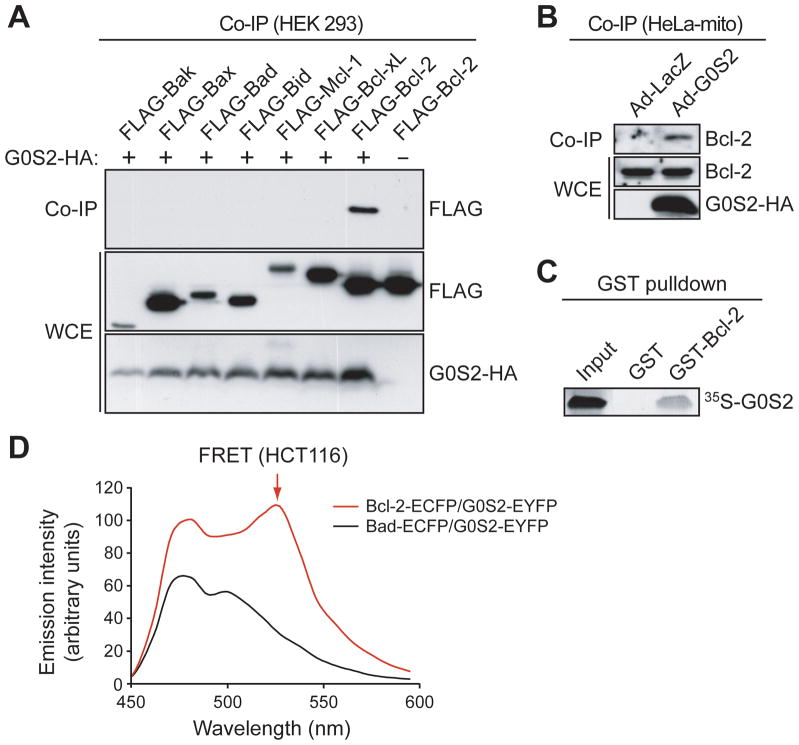
Figure 2.
G0S2 directly interacts with the anti-apoptotic protein Bcl-2. A, HEK293 cells were co-transfected with plasmids expressing G0S2-HA and a FLAG-tagged Bcl-2 protein. G0S2 was immunoprecipitated using an α-HA antibody, and the immunoprecipitate was analyzed by immunoblotting with an α-FLAG antibody. The presence G0S2 and Bcl-2 member were also monitored in the whole cell extract (WCE). B, G0S2 was immunoprecipitated from the mitochondrial fraction of Ad-G0S2- or Ad-LacZ-infected HeLa cells using an α-HA antibody, and the immunoprecipitate was analyzed using an α-Bcl-2 antibody. C, GST pull-down assays using purified GST-tagged Bcl-2 and 35S-methionine labeled G0S2. D, FRET analysis. Fluorescence emission spectra in HCT116 cells co-expressing G0S2-EYFP and either Bcl-2-ECFP or Bad-ECFP. The peak of fluorescence emission at 525 nm (FRET signal) is indicated by the arrow.

