
Fig. 2.
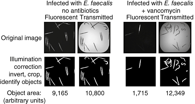
Automated high-throughput Caenorhabditis elegans curing assay in 384-well plates. Image analysis quantifies worm survival. Top row shows raw fluorescent SYTOX orange and bright field images captured from an untreated well and an antibiotic-treated well. The images were analysed using the image analysis software CellProfiler through a pipeline of 29 processing steps, which is shown in the bottom row. The total object area of the fluorescent and the bright field images are measured. The areas of the SYTOX orange objects and bright field objects are used to approximate the number of dead worms and the total number of worms, respectively. Worm survival is calculated as 1-(SYTOX orange area/total bright field worm area).