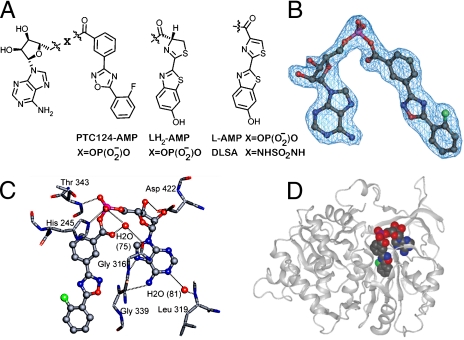
Fig. 1.
Chemical and cocrystal structures. (A) Comparison of the structure of the mixed anhydrides of AMP (Left) with PTC124, D- luciferin (LH2), dehydroluciferin (L), and DLSA. (B) Fo-Fc omit map of the PTC124-AMP mixed anhydride contoured at 3σ. (C) Hydrogen-bonded interactions between PTC124-AMP MAI (6, ball and stick) and luciferase residue (sticks) are indicated as dashed lines. (D) Cocrystal X-ray structure of FLuc and PTC124-AMP MAI.

