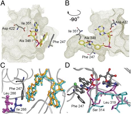
Fig. 2.
Substrate binding pocket of FLuc with PTC124-AMP bound. (A, B) Two pocket orientations. (C) Structure of FLuc (Gray Ribbon) liganded with PTC124-AMP (Turquoise) overlaid on the structure of DLSA (Gold) bound to LcrLuc (PDB: 2D1S) showing the close similarity of ligand binding modes. Leu286 in FLuc corresponds to Ile288 in LcrLuc. These residues are located at the end of the respective ligand binding pockets, but whereas Ile288 moves upon ligand binding, Leu286 does not. (D) Overlay of the structures of apo-FLuc (Magenta) and FLuc liganded with the PTC124-AMP MAI adduct (6, Turquoise) showing movement of a loop between residues Ser314 and Leu319 upon binding of ligand (ball and stick).

