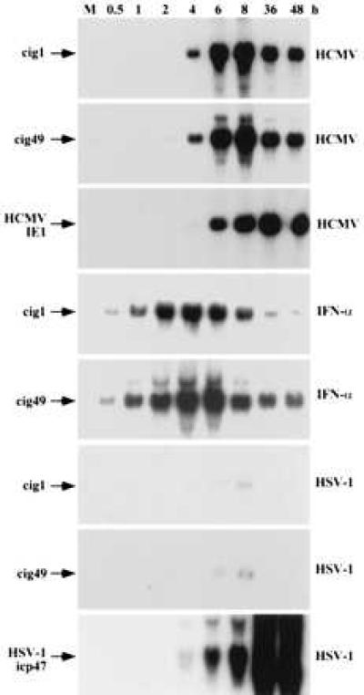
Figure 5.

Kinetic analysis of cig RNA accumulation. HF cells were mock-infected (M) or treated with the inducers identified to the right (HCMV, HSV-1, and IFN-α), RNA was prepared at various times after treatment (indicated above lanes), and analyzed by Northern blot by using the probes indicated on the left (cig1, cig49, HCMV IE1, and HSV-1 icp47).
