Fig. 3.
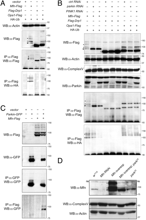
Mfn is ubiquitinated in a PINK1/Parkin-dependent manner in vitro and in vivo. (A) S2R+ cells cotransfected with HA-Ub plus empty vector or Flag-tagged Mfn, Drp1, or Opa1 plasmids as indicated were immunoprecipitated and subjected to Western blot analysis. (B) S2R+ cells were treated with control, parkin, and PINK1 RNAi before being transfected as indicated. Cells were harvested and subjected to Western blot analysis as shown. Samples were also subjected to immunoprecipitation, and Western bots were probed with antibodies against Flag and HA. (C) Immunoprecipitates of S2R+ cells expressing combinations of empty vector, parkin-GFP, and Mfn-Flag as shown. (D) Western blot analysis of Drosophila Mfn levels in vivo. A nonspecific band seen in all samples is denoted “ns.” Complex Vα and actin are used as loading controls. Genotypes: w1118, wild type; Mfn RNAi, da-GAL4,UAS-Mfn-RNAi; Mfn overexpression, da-GAL4,UAS-Mfn; Mfn overexpression park25, da-GAL4,UAS-Mfn,park25/park25; park25, park25/park25. In all panels, black arrowheads indicate full-length Mfn, asterisks are ubiquitinated Mfn, white arrowheads denote full-length Opa1, white diamonds are ubiquitinated Opa1, and white arrows show full-length Drp1.

