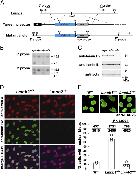
Fig. 1.
Inactivation of mouse Lmnb2. (A) Gene-targeting strategy to replace the coding sequences of Lmnb2 exon 1 with a lacZ cassette, beginning at the translation initiation site (ATG). The positions of relevant EcoRI and EcoRV sites and the 5′, 3′, and neo probes for Southern blot analysis are shown. (B) Southern blots identifying gene-targeting events in mouse embryonic stem cells. Genomic DNA was digested with EcoRV. (C) Western blots of mouse embryonic fibroblast extracts with antibodies against lamin B1 and lamin B2. Actin was used as a loading control. (D) Immunofluorescence microscopy of Lmnb2+/+ and Lmnb2−/− embryonic fibroblasts with antibodies against lamin A and lamin B2. (Bottom) Merged images with DAPI staining for nuclear DNA. (E) Lmnb2−/− fibroblasts did not have a higher frequency of cells with nuclear blebs than WT cells. (Upper) Confocal images of Lmnb2+/+, Lmnb1−/−, and Lmnb2−/− fibroblasts stained with a monoclonal antibody against LAP2β, a protein of the inner nuclear membrane. (Lower) graph showing the percentage of cells with nuclear blebs. Each open circle represents an independent cell line (two WT, three Lmnb1−/−, and four Lmnb2−/− cell lines). The ratio above each bar indicates the number of cells with nuclear blebs over the total number of cells evaluated.

