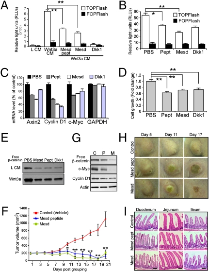
Fig. 5.
Therapeutic effect of Mesd treatment on human breast cancer cells and MMTV-Wnt1 tumor xenografts. (A) Wnt inhibitory effect was analyzed by measuring luciferase activity in Wnt3a-stimulated HEK293 cells stably expressing TOPFlash reporter treated with indicated reagents. Cells were incubated with L CM, Wnt3a CM, or Wnt3a CM together with Mesd protein (1 μM), Mesd peptides (1 μM) or Dkk1 (10 nM) for 16 h at 37°C. (B and C) Treatment with Mesd (5 μM), Mesd peptide (5 μM), or Dkk1 (50 nM) decreased Wnt signaling in unstimulated HCC1187 cells examined by TOPFlash reporter assay (B) and Wnt target gene expression assessed by qRT-PCR (C). (D and E) Mesd treatment suppressed free β-catenin accumulation (E) and cell growth (D) in HCC1187 cells. (F) Mesd and its peptide significantly inhibited tumor growth. Representative pictures of tumors upon treatment are shown in H. Mice bearing established MMTV-Wnt1 tumor transplants were divided into three groups (five animals per group) and were i.p. injected with PBS (vehicle), Mesd, or Mesd peptide every other day. Tumor volume was analyzed using caliper measurement. Data represent at least three independent experiments; each time point represents the mean tumor volume ± SEM. *P < 0.05; **P < 0.01. (G) Mesd peptide (P) and Mesd (M) treatment decreased Wnt signaling compared with control (C) treatment as confirmed by GST–E-cadherin pull-down and target gene expressions in MMTV-Wnt1 tumors. (I) No significant adverse effect on small intestine with Mesd and Mesd peptide administration is apparent upon gross examination. (Scale bar, 50 μm.)

